Past Issues
Phakomatosis Pigmentovascularis Type Va: A Newborn Case
Mustafa Yildiz1, Samet Benli2,*
1Department of Pediatrics, Elazig Fethi Sekin City Hospital Elazig, Turkey
2Department of Pediatrics-Neonatology, Cengiz Gökçek Gynaecology and Paediatrics Hospital Gaziantep, Turkey
*Corresponding author: Samet Benli, Department of Pediatrics-Neonatology, Cengiz Gökçek Gynaecology and Paediatrics Hospital, 27000, Gaziantep, Turkey, E-mail: [email protected]
Received Date: March 28, 2024
Published Date: June 13, 2024
Citation: Yildiz M, et al. (2024). Phakomatosis Pigmentovascularis Type Va: A Newborn Case. Neonatal. 5(2):18.
Copyrights: Yildiz M, et al. © (2024).
ABSTRACT
Phacomatosis pigmentovascularis (PPV) is characterised by a combination of vascular and pigmentary birthmarks. The association of cutis marmorata telangiectatica congenita with aberrant dermal melanocytosis has been named phacomatosis pigmentovascularis type V. Approximately 50% of patients have ocular, neurological or skeletal involvement, which requires further investigation. Accurate and early diagnosis will be important for the treatment of the patient.
Keywords: Phacomatosis Pigmentovascularis, Pediatrician, Type V, Cutis Marmorata Telangiectatica
INTRODUCTION
Phakomatosis pigmentovascularis (PPV) is a rare congenital malformation syndrome characterised by a combination of capillary abnormalities and dermal melanocytosis present from birth and classified into 4 types by Hasefawa and Yasuhara according to the different characteristics of vascular and pigmentary malformations. This classification is divided into 2 subtypes according to the absence (type a) or presence (type b) of extracutaneous involvement. In 2003, Torrelo et al. described cutismarmorata telangiectatica congenita (CMTC) associated with abnormal Mongolian spots and PPV type V [1-3]. Phacomatosis pigmentovascularis (PPV) is a rare congenital disease characterised by the coexistence of vascular and pigmentary nevi (MS, nevus of Ota, verrucous nevus and nevus spilus) with or without extracutaneous manifestations [4]. When the number of PPV type V cases was reviewed in the literature, a small number of cases were found. We wanted to present a twenty-day-old patient with phakomatosis pigmentovascularis (PPV) type Va who had not been previously diagnosed.
CASE REPORT
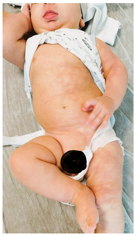
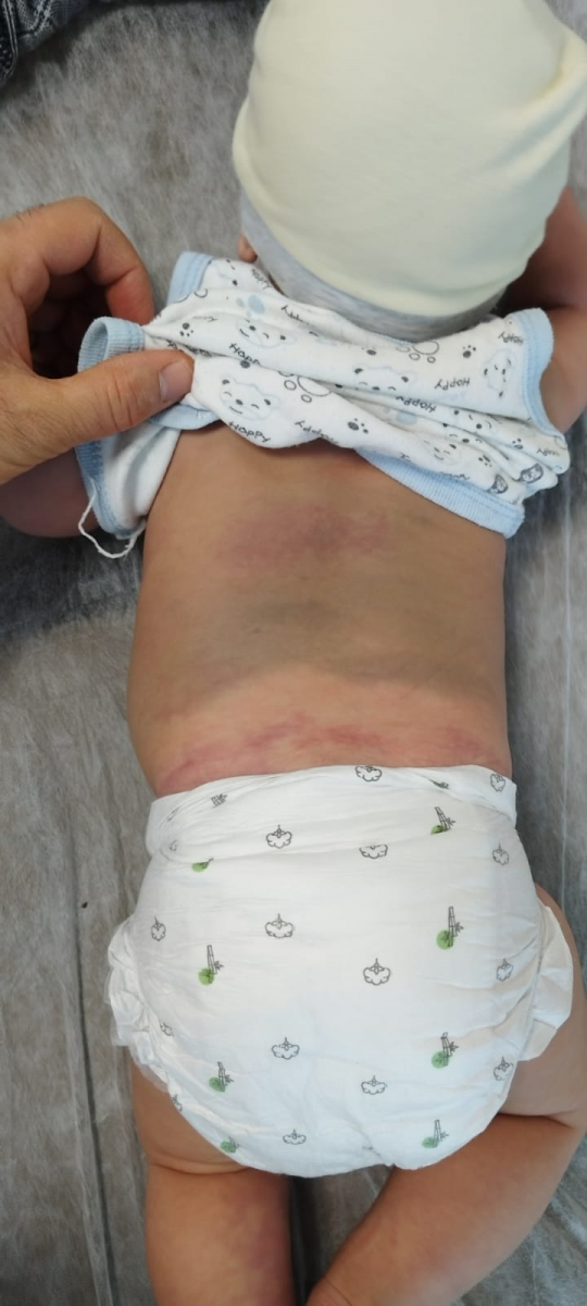
The patient, born by caesarean section at thirty-eight weeks of gestation from the fourth pregnancy of a 34-year-old mother, had no abnormality in antenatal follow-up. The patient who presented to the paediatric outpatient clinic because of skin findings present since birth was admitted to our unit for investigation and treatment. His body weight was 4350 grams (50-75p), height 54 cm (25p) and head circumference 37.8 (25-50p). Skin examination findings of the patient revealed cutis marmorata telangiectatica congenita on the left lower extremity, cutis marmorata telangiectatica congenita appearance starting from below the left nipple on the left half of the body, mongol spot on the back, diffuse cutis marmorata telangiectatica congenita on the back and diffuse haemangioma on the scalp (Figure 1a-1b). Phakomatosis pigmentovascularis (PPV) type V was considered with physical examination findings. Ophthalmological examination was performed in terms of eye pathologies and was normal. Abdominal, urinary and cranial ultrasonography revealed no abnormality. Echocardiography did not reveal any pathological condition. Brain magnetic resonance imaging (MRI) performed to evaluate central nervous system pathologies was reported normal. A skin biopsy was planned. It could not be performed because the family did not give consent. The patient was followed up by dermatology and paediatrics with a diagnosis of PPV type Va.
Figure 1a. Appearance of cutis marmorata telangiectatica congenita starting under the left nipple on the left half of the body
Figure 1b. Mongolian spot on the back and cutis marmorata telangiectatica congenita
DISCUSSION
Phacomatosis pigmentovascularis (PPV) has been defined as the clinical association of dermal melanocytic nevus and vascular malformation. PPV is classified into five types according to epidermal findings and vascular malformation (Table 1) [5].
Table 1. Present classification of distinct types of phacomatosis pigmentovascularis
|
Type |
Nevus Findings |
Pigmentary Findings |
|
I |
Nevus flammeus |
Nevus pigmentosus |
|
II |
Nevus flammeus and/or anemic nevus |
Mongolian spot
|
|
III |
Nevus flammeus and/or anemic nevus |
Nevus spilus
|
|
IV |
Nevus flammeus and/or anemic nevus |
Mongolian spot, nevus spilus
|
|
V |
Cutis marmorata telangiectatica congenita
|
Mongolian spot |
Each type is classified as type a if only oculocutaneous disease is present and type b if extracutaneous disease is present. The association of dermal melanocytosis with cutis marmorata telangiectatica congenita (CMTC) was first described by Enjolras and Mulliken. This association was found in two more patients and PPV type V defined this association as a separate classification [2].
Cutis marmorata telangiectatica congenital is observed only in PPV type V. Cutis marmorata telangiectatica congenita (CMTC) is a rare congenital disease in which telangiectasia, phlebectasia, skin atrophy and ulceration may be observed. Although the etiology is not known exactly, it is thought to be multifactorial. Associated anomalies are present in over 50% of cases. Neurological, musculoskeletal, ophthalmological, cardiovascular and cutaneous abnormalities have been associated with CMTC. Other cutaneous findings observed in patients include nevus flammeus, haemangioma and café-au-lait spots [5,6]. There are many diseases reported to be associated with PPV. Right kidney agenesis, scoliosis, multiple granular cell tumours, Sturge-Weber syndrome, colonic polyposis, CTS, multiple renal angiomatosis, leg length discrepancy, hypoplastic larynx, subglottic stenosis, epilepsy, hypoplasia of portal veins, Cerebral atrophy, iris mamilations, hypoplasia of the inferior vena cava, iliac and femoral veins, iris hamartomas, glaucoma, multiple granular cell tumours and congenital triangular alopecia are among the diseases [7-9]. In our case, no extracutaneous disease was present.
The exact aetiology of PPV is unknown. The genetic phenomenon of non-allelic twin spotting has been proposed as a mechanism to explain the association between CMTC and dermal melanocytosis . This is a concept in which two different mutant alleles at the same locus or nearby loci (via somatic recombination) create two genetically distinct clones of neighbouring mutant cells in a background of normal cells [10].
PPV type V cases are rare in the literature and the diagnosis is usually clinical. PPV without systematic complications is benign and does not require treatment. Ocular abnormalities, especially melanosis oculi, are common in PPV type V. An eye examination by an ophthalmologist is recommended for patients with the diagnosis. Patients with CMTC should be evaluated for associated anomalies, especially body asymmetry, glaucoma and neurological disorders.
CONCLUSION
Phakomatosis pigmentovascularis is a benign clinical picture without visceral involvement. In case of organ involvement, the patient should be followed up in a multidisciplinary approach.
FUNDING
The authors received no financial support for the research, authorship, and/or publication of this article.
CONFLICT OF INTEREST
The authors declare that there is no any conflict of interest.
REFERENCES
- Hasegawa Y, Yasuhara M. (1985). Phakomatosis pigmentovascularis type IVa. Arch Dermatol. 121(5):651-655.
- Torrelo A, Zambrano A, Happle R. Cutis marmorata telangiectatica congenita and extensive mongolian spots: type 5 phacomatosis pigmentovascularis. Br J Dermatol. 2003 Feb;148(2):342-345.
- Happle R. (2005). Phacomatosis pigmentovascularis revisited and reclassified. Arch Dermatol. 141(3):385-388.
- Byrom L, Surjana D, Yoong C, Zappala T. (2015). Red-white and blue baby: a case of phacomatosis pigmentovascularis type V. Dermatol Online J. 21(6):13030/qt2b0980p8.
- Ha JW, Hahm JE, Park SE, Lee JY, Kim CW, Kim SS. (2017). A Case of Phacomatosis Pigmentovascularis Type IIa in a Korean Infant. Ann Dermatol. 29(5):638-639.
- Amitai DB, Fichman S, Merlob P, Morad Y, Lapidoth M, Metzker A. (2000). Cutis marmorata telangiectatica congenita: clinical findings in 85 patients. Pediatr Dermatol. 17(2):100-104.
- Shields CL, Kligman BE, Suriano M, Viloria V, Iturralde JC, Shields MV, et al. (2011). Phacomatosis pigmentovascularis of cesioflammea type in 7 patients: combination of ocular pigmentation (melanocytosis or melanosis) and nevus flammeus with risk for melanoma. Arch Ophthalmol. 129(6):746-750.
- Dutta A, Ghosh SK, Bandyopadhyay D, Bhanja DB, Biswas SK. (2019). Phakomatosis Pigmentovascularis: A Clinical Profile of 11 Indian Patients. Indian J Dermatol. 64(3):217-223.
- Seckin D, Yucelten D, Aytug A, Demirkesen C. (2007). Phacomatosis pigmentovascularis type IIIb. Int J Dermatol. 46(9):960-963.
- Devillers AC, de Waard-van der Spek FB, Oranje AP. (1999). Cutis marmorata telangiectatica congenita: clinical features in 35 cases. Arch Dermatol. 135(1):34-38.
 Abstract
  PDF
PDF
